With FPG Pulmonologist Omar Sheriff, MD
Lung cancer is the leading cause of cancer-related deaths in the entire world, with the highest mortality rate of all cancers, for both men and women. Unfortunately, it’s also one of the hardest cancers to diagnose, with symptoms not appearing until the malignancy has progressed and the lungs being notoriously difficult to biopsy.
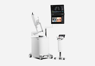
But with the introduction of the Ion endoluminal system, a robotic-assisted platform for minimally invasive biopsy in the lungs, physicians at Sarasota Memorial are using state-of-the-art technology to tackle those difficult cases and diagnose lung cancer earlier.
“And if we can diagnose lung cancer at its earliest stages, patient survival is the greatest,” says Dr. Omar Sheriff. “That’s why this is so important.”
Screening for Lung Cancer?
Guidelines for lung cancer screening have recently changed.
Click here to read more and see if you should be talking to you doctor about scheduling a screening.
A New Gold Standard
For decades, the gold standard in lung biopsy technique was startlingly direct: using a CT scan as guidance, a physician would insert a needle into the patient’s chest, piercing the chest wall and then, hopefully, contacting the lung nodule or lesion of interest. But while the CT-guided needle biopsy proved an effective method for most cases, complication rates were high, with the risk of lung collapse at 20%, and smaller or deeper lung nodules were often inaccessible.
Even the advent of robotic-assisted platforms for minimally invasive biopsies ran into roadblocks, with early systems lowering complications drastically but returning less definitive results. And every method encountered the same recurring problem: the patient needs to breathe during the procedure, meaning the lungs are inflating and deflating as the surgeon is conducting the biopsy. “And suddenly, the lung nodule becomes a moving target,” says Dr. Sheriff. And a lung nodule that was less than 1cm in diameter on the CT scan can be shifting as much as 2cm with each breath. “That leaves a lot of room for error,” he says. “So we need precise tech to let us know we’re in the right location.”
Enter the Ion endoluminal system, setting the new gold standard.
How Does The Ion Endoluminal System Work?
Like previous platforms for robotic-assisted minimally invasive lung biopsies, the process begins with a CT scan of the lungs and chest. Captured in high resolution, this data is uploaded to the Ion system, which converts the scan into a high-definition, high-detail ‘roadmap’ of the patient’s lungs that includes all of the little nooks and crannies. From there, the physician can identify whatever nodule or lesion is of interest and the Ion system will automatically plot the most efficient course to get there. “Like putting an address into a GPS,” says Dr. Sheriff.
This information then feeds directly into a special endoscope equipped with shape-sensing technology—millions of tiny hair-like optical fibers embedded into the scope—that is able to use the Ion roadmap to guide the catheter through the labyrinthine lobes of the lungs and to the predesignated biopsy target, even as the lungs move. “The system gives us information and feedback in real time,” says Dr. Sheriff, “so we know exactly where that catheter is.” And at only 3.5mm in diameter, the catheter is more flexible than previous models, articulating 180 degrees in every direction, enabling it to access even hard-to-reach nodules near the lung lining.
Perhaps more importantly, the Ion system means physicians can access the more central lung nodules for biopsy, potentially allowing them to diagnose lung cancer even earlier than before. “And that’s something you just cannot do with a CT-guided needle biopsy,” says Dr. Sheriff.
Impacts In Real Time
According to Dr. Sheriff, patients and physicians using the Ion system are already seeing a difference, including a “significant increase” in diagnosis of bronchial lung nodules. “Close to 90%,” says Dr. Sheriff, and the majority have been of a size that could easily have confounded earlier biopsy techniques.
And with Ion already testing new technology that allows the system to interface with CT scans taken in real-time, during the procedure, physicians will soon be able to make micro-adjustments in the moment, Dr. Sheriff says, perfectly melding machine precision with human intuition.
Learn More
To learn more about lung cancer screening and diagnostics at the Sarasota Memorial Brian D. Jellison Cancer Institute, click here.
 Omar Sheriff, MD is a board certified pulmonology and critical care physician with First Physicians Group. For more information or to make an appointment, click here.


